Retina: Exploring Structure, Function, and Innovations
Abstract: The retina, a complex neural tissue lining the posterior segment of the eye, plays a pivotal role in vision by converting light into neural signals that are transmitted to the brain. This article provides a comprehensive overview of the retina, covering its anatomy, physiology, pathology, and recent advancements in diagnostic and therapeutic modalities. By elucidating the intricacies of retinal structure and function, ophthalmologists can enhance their understanding of retinal diseases and refine their management strategies to optimize patient outcomes.
Introduction: The retina serves as the sensory receptor for vision, orchestrating the initial stages of visual processing before transmitting signals to higher visual centers in the brain. Understanding the structure, function, and pathophysiology of the retina is essential for diagnosing and managing a diverse array of retinal diseases.
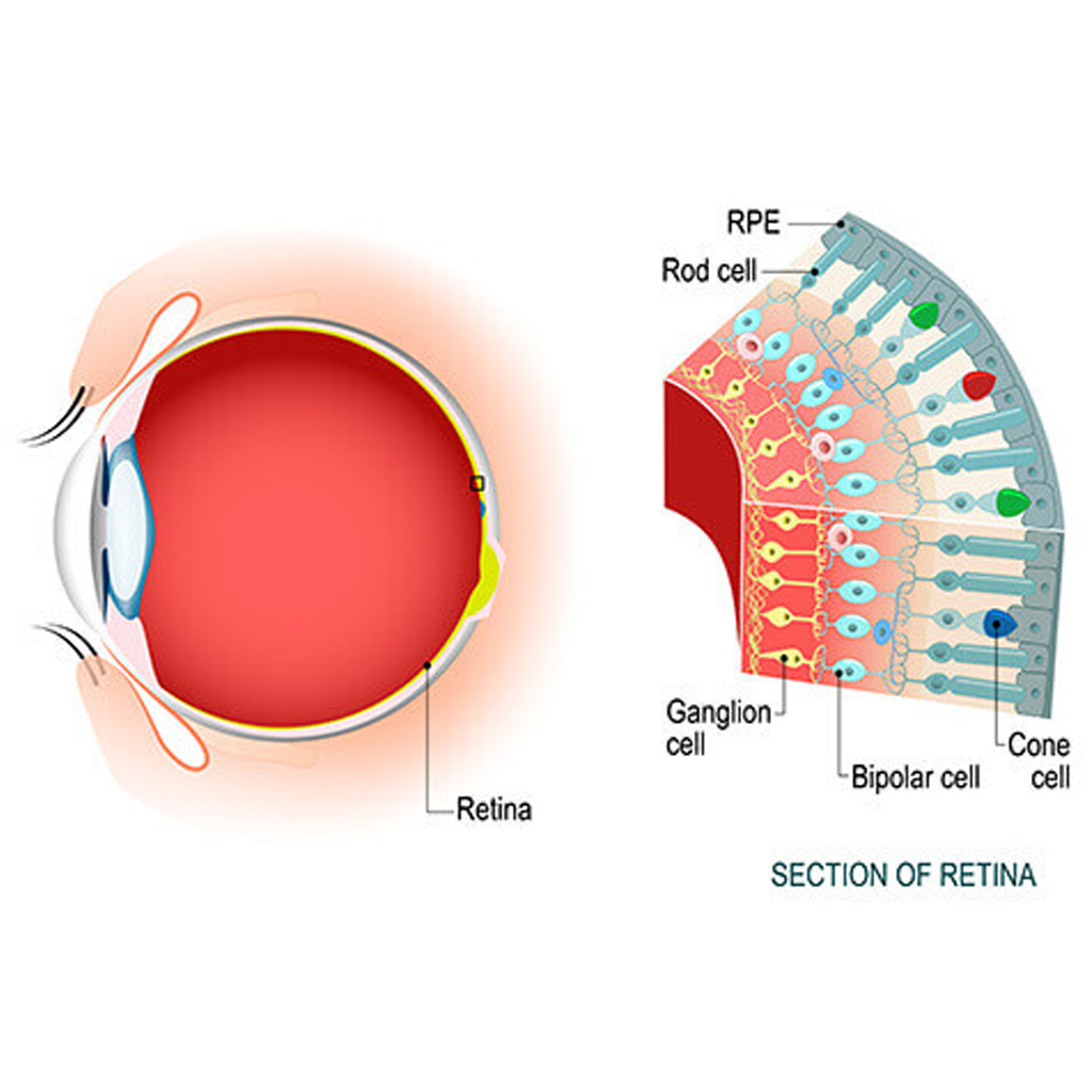
Anatomy of the Retina: The retina consists of multiple layers, including photoreceptor cells (rods and cones), bipolar cells, ganglion cells, and various interneurons. Specialized regions such as the macula and fovea centralis harbor a high density of cones, facilitating detailed central vision.
Physiology of Vision: Visual transduction begins in the photoreceptor cells, where light activates photopigments, initiating a cascade of biochemical events that culminate in the generation of neural signals. These signals are then transmitted through the retinal layers to the optic nerve head and subsequently to the visual cortex via the optic nerve.
Retinal Diseases: Retinal diseases encompass a broad spectrum of conditions ranging from age-related macular degeneration (AMD) and diabetic retinopathy to retinal detachment and inherited retinal dystrophies. Each condition presents unique challenges in diagnosis and management, necessitating a tailored approach based on disease severity and patient-specific factors.
Diagnostic Modalities: Advances in imaging technologies have revolutionized the diagnosis and monitoring of retinal diseases. Optical coherence tomography (OCT), fundus autofluorescence (FAF), fluorescein angiography (FA), and indocyanine green angiography (ICGA) provide detailed insights into retinal structure, function, and vascular dynamics, enabling precise disease characterization.
Therapeutic Interventions: Treatment options for retinal diseases have expanded significantly in recent years, with intravitreal pharmacotherapy (e.g., anti-vascular endothelial growth factor agents, corticosteroids) revolutionizing the management of conditions such as AMD and diabetic macular edema. Surgical interventions, including vitrectomy and retinal laser procedures, remain essential for addressing retinal detachment and proliferative vitreoretinopathy.
Emerging Innovations: Ongoing research efforts are focused on developing novel therapies targeting specific molecular pathways implicated in retinal disease pathogenesis. Gene therapy, stem cell transplantation, and retinal prosthetic devices offer promising avenues for restoring vision in degenerative retinal disorders.
Conclusion: The retina serves as a window to the visual world, and its intricate structure and function underlie the foundation of vision. By staying abreast of the latest innovations in retinal imaging, diagnostics, and therapeutics, ophthalmologists can provide comprehensive care to patients with retinal diseases, preserving vision and improving quality of life.
For further reading and reference:
- American Academy of Ophthalmology – Retina/Vitreous: https://www.aao.org/bcscsnippetdetail.aspx?id=58c279e0-9799-4765-b456-19cd852740d0
- Retina Society – Patient Information: https://www.retinasociety.org/patient-information


