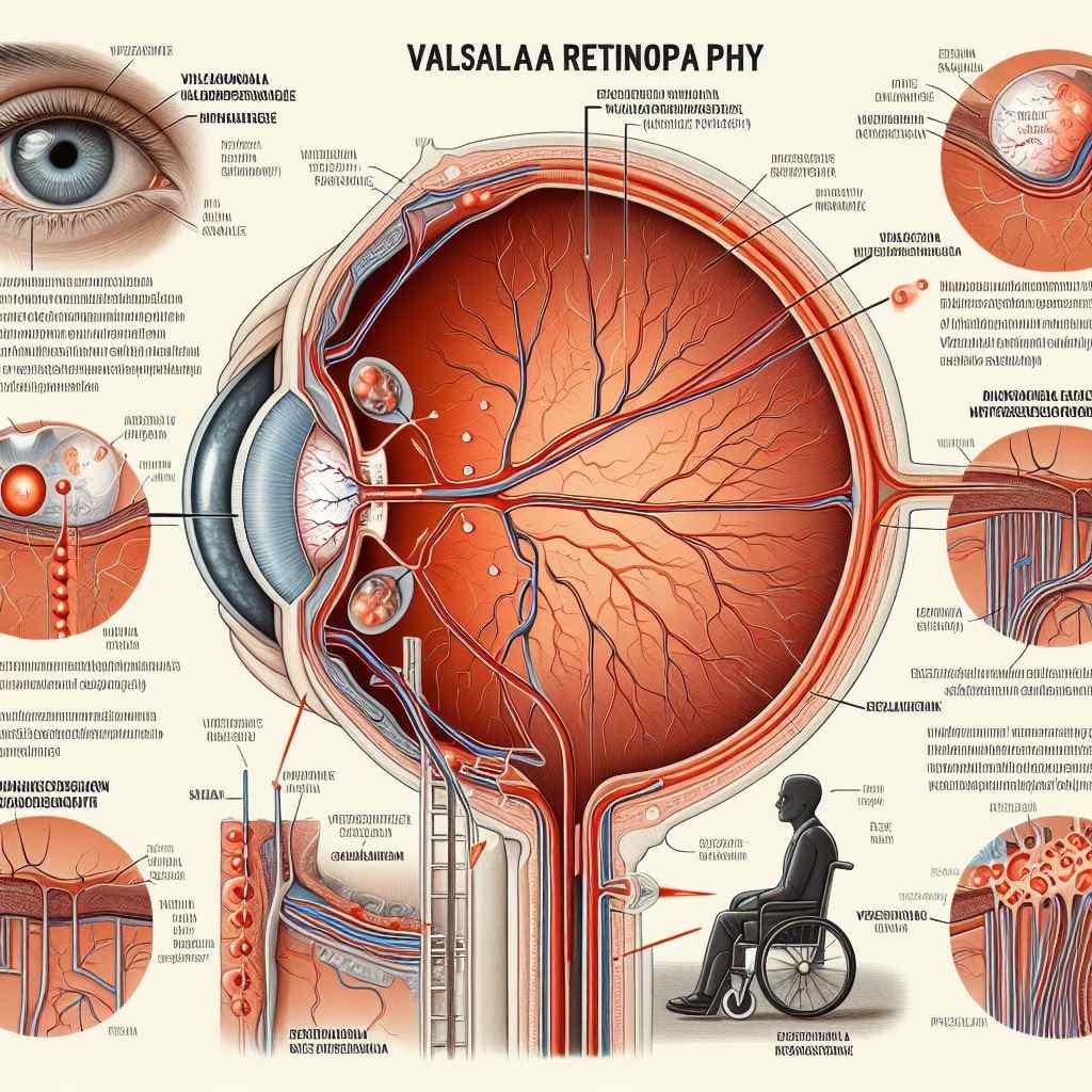
Exploring Valsalva Retinopathy: Understanding Causes, Symptoms, and Treatment
Introduction: Valsalva retinopathy is a condition characterized by bleeding into the retina due to a sudden increase in intraocular pressure following a Valsalva maneuver. This article provides an in-depth exploration of Valsalva retinopathy, including its pathophysiology, clinical presentation, diagnostic considerations, and treatment options.
Pathophysiology: Valsalva retinopathy occurs when a sudden increase in intraocular pressure leads to the rupture of superficial retinal vessels, resulting in hemorrhage within the retina. The Valsalva maneuver, which involves forced expiration against a closed airway (e.g., during coughing, vomiting, or weightlifting), causes a transient increase in intrathoracic and intra-abdominal pressure, subsequently raising intraocular pressure. The sudden rise in pressure can exceed the retinal venous pressure, leading to vessel rupture and bleeding.
Clinical Presentation: Patients with Valsalva retinopathy typically present with sudden, painless vision loss or visual disturbances following a Valsalva maneuver. Clinical features may include:
- Floaters or spots in the visual field.
- Blurred vision or vision loss, which may be transient or persistent.
- Fundoscopic examination revealing preretinal or intraretinal hemorrhages, often in the macular region.
Diagnostic Considerations: Diagnosis of Valsalva retinopathy is based on clinical history, symptoms, and fundoscopic findings. Diagnostic considerations include:
- Detailed history regarding recent Valsalva-like activities or events.
- Ophthalmic examination, including dilated fundus examination, to visualize retinal hemorrhages and assess macular involvement.
- Ancillary tests such as optical coherence tomography (OCT) to evaluate the extent of macular involvement and rule out associated macular pathology.
Treatment Options: Management of Valsalva retinopathy depends on the extent and severity of retinal hemorrhage and associated symptoms. Treatment options may include:
- Observation: Small, asymptomatic hemorrhages may resolve spontaneously over time without intervention.
- Pars plana vitrectomy: Surgical intervention may be considered for large or visually significant hemorrhages, particularly those involving the macula. Vitrectomy allows for the removal of blood from the vitreous cavity and facilitates rapid visual recovery.
- Anti-VEGF therapy: Intravitreal injection of anti-vascular endothelial growth factor (VEGF) agents may be beneficial in cases with persistent macular edema or neovascularization.
Prognosis: The prognosis of Valsalva retinopathy is generally favorable, with many cases experiencing spontaneous resolution of retinal hemorrhages and improvement in visual symptoms over time. However, the extent of visual recovery may vary depending on the severity of macular involvement and the presence of associated complications such as macular fibrosis or tractional retinal detachment.
Reference Sites:
- American Academy of Ophthalmology (AAO) – https://www.aao.org/
- National Eye Institute (NEI) – https://www.nei.nih.gov/
- PubMed Central (PMC) – [Link to relevant research articles and case studies]
By providing a comprehensive overview of Valsalva retinopathy, this article aims to enhance awareness among healthcare professionals and improve the management of this condition. Continued research efforts are needed to further elucidate the pathophysiology and optimize treatment strategies for affected individuals.


