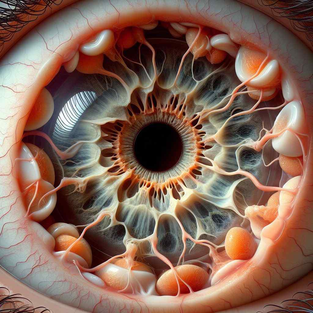
Exploring Congenital Optic Disc Anomalies: Insights into Classification, Clinical Features, Diagnosis, Management, and Recent Advances
- By Anshu Dagar
Abstract: Congenital optic disc anomalies encompass a spectrum of structural variations affecting the optic nerve head, presenting unique challenges in diagnosis and management. This article provides a comprehensive review of congenital optic disc anomalies, covering their classification, clinical manifestations, diagnostic approaches, treatment strategies, and recent innovations. By elucidating the complexities of these anomalies, ophthalmologists can enhance their diagnostic acumen and optimize patient care through tailored interventions.
Introduction: Congenital optic disc anomalies comprise a diverse array of developmental abnormalities affecting the optic nerve head. Understanding the spectrum of anomalies and their associated clinical features is essential for accurate diagnosis and appropriate management.
Classification: Congenital optic disc anomalies are classified based on their morphological features, including optic disc coloboma, optic disc hypoplasia, morning glory disc anomaly, and optic disc pit. Each subtype exhibits distinct characteristics and may be associated with varying degrees of visual impairment.
Clinical Features: Clinical presentation of congenital optic disc anomalies varies depending on the specific subtype and severity of the anomaly. Common features include optic nerve head excavation, vessel displacement, retinal pigmentary changes, and visual field defects.
Diagnostic Modalities: Diagnosis of congenital optic disc anomalies requires a thorough ophthalmic examination, including fundoscopy, optical coherence tomography (OCT), and visual field testing. Additional imaging modalities such as fluorescein angiography and magnetic resonance imaging (MRI) may aid in delineating the extent of optic nerve involvement.
Management Strategies: Management of congenital optic disc anomalies focuses on preserving visual function and addressing associated complications such as amblyopia and refractive errors. Treatment modalities may include optical correction, visual rehabilitation, and surgical interventions for specific indications.
Recent Advances: Recent advancements in the field of congenital optic disc anomalies include novel imaging techniques such as OCT angiography (OCT-A) for better visualization of vascular abnormalities, genetic testing for identifying underlying etiologies, and emerging surgical approaches for complex cases.
Conclusion: Congenital optic disc anomalies present a diagnostic and therapeutic challenge in ophthalmology, requiring a nuanced understanding of their classification, clinical features, and management strategies. By staying informed about the latest research and innovations in this field, ophthalmologists can enhance their ability to diagnose and manage congenital optic disc anomalies effectively, ultimately improving visual outcomes for affected individuals.
For further reading and reference:
- American Academy of Ophthalmology – Optic Disc Anomalies: https://www.aao.org/bcscsnippetdetail.aspx?id=d6453b50-7db5-4a6b-9b4c-b94e59f89a1e
- Journal of Pediatric Ophthalmology & Strabismus – Congenital Optic Disc Anomalies: https://journals.lww.com/jposna/pages/default.aspx
- National Institutes of Health – Congenital Optic Disc Anomalies: https://rarediseases.org/rare-diseases/optic-disc-anomalies-congenital/


