Understanding Choroidal Nevi: A Comprehensive Overview
Choroidal nevi, although generally benign, present diagnostic and management challenges due to their potential for malignant transformation and associated vision-threatening complications. This article aims to provide a thorough exploration of choroidal nevi, encompassing their epidemiology, clinical features, diagnostic evaluation, management strategies, and recent advancements in the field.
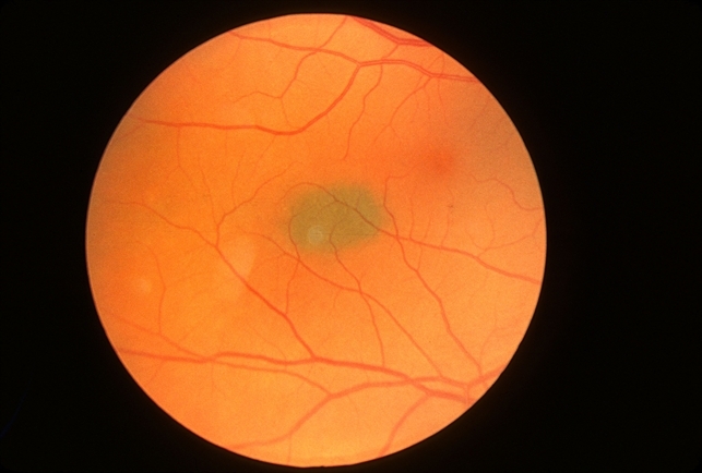
Introduction: Choroidal nevi are common intraocular lesions composed of benign melanocytic cells. While often asymptomatic and incidentally discovered during routine eye examinations, choroidal nevi require careful evaluation and monitoring to differentiate them from malignant counterparts and detect potential transformation.
Epidemiology: Choroidal nevi are prevalent in the general population, with estimates suggesting that up to 5-10% of adults harbor these lesions. They typically manifest in individuals over 40 years of age and are more frequently observed in Caucasians. Despite their high prevalence, only a small percentage of choroidal nevi undergo malignant transformation.
Clinical Features: Choroidal nevi typically present as well-defined, pigmented lesions located within the choroid. They may exhibit a variety of characteristics on clinical examination, including flat or slightly elevated morphology, uniform or variegated pigmentation, and associated drusen or overlying subretinal fluid. While most choroidal nevi remain stable over time, changes in size, shape, or associated symptoms warrant further evaluation.
Diagnostic Evaluation: Accurate diagnosis and risk stratification of choroidal nevi rely on a combination of clinical assessment and ancillary diagnostic studies. Multimodal imaging modalities such as fundus photography, optical coherence tomography (OCT), fundus autofluorescence, and ultrasonography play a pivotal role in characterizing lesion morphology, documenting growth patterns, and identifying features suggestive of malignant transformation.
Management Strategies: The management of choroidal nevi entails a judicious balance between vigilant observation and targeted intervention. Factors influencing treatment decisions include lesion size, location, associated symptoms, and patient preferences. While most choroidal nevi do not require active intervention, close monitoring at regular intervals is recommended to detect potential signs of malignancy or vision-threatening complications.
Recent Advancements: Recent advancements in imaging technology and molecular diagnostics have enhanced our understanding of choroidal nevi and improved risk stratification algorithms. Emerging techniques such as enhanced depth imaging OCT, wide-field imaging, and molecular genetic profiling hold promise in predicting the likelihood of malignant transformation and refining personalized management approaches.
Conclusion: In conclusion, choroidal nevi represent a common yet diagnostically challenging entity in clinical ophthalmology. Through a comprehensive understanding of their clinical features, diligent diagnostic evaluation, and evidence-based management strategies, ophthalmologists can effectively navigate the complexities of choroidal nevi and optimize patient outcomes.
Links: